The Melbourne Brain Centre Imaging Unit (MBCIU) offers human medical imaging using PET/CT and ultrahigh-field MRI. We scan human participants in research trials to investigate disease. We also scan animals, plants, medical implants and artefacts.
The MBCIU offers two types of imaging: magnetic resonance imaging (MRI) and positron emission tomography combined with computed tomography (PET/CT).
We work with researchers and clinicians to study diseases and normal biological processes. Our diagnostic imaging technologists and academics have backgrounds in imaging physics, neuroscience and engineering. We have expertise in anatomical, functional and molecular brain imaging. We also have expertise imaging other areas of the body, such as the eye and orbit, the spine, and the musculoskeletal system.
The MBCIU team works with scientists and engineers who require non-destructive internal and external imaging. We can perform imaging of animal specimens, fossils, museum artefacts, materials and equipment.
MRI
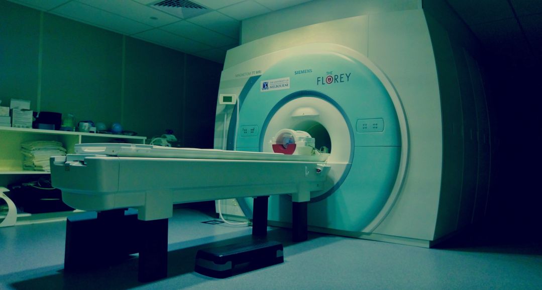
The MBCIU has a 7 Tesla (7T) MRI scanner – one of only two of its kind in Australia. This ultrahigh-field scanner acquires structural, functional and molecular information. Imaging is non-invasive and at high spatial and temporal resolutions.
The 7T MRI scanner is used in a range of research studies. In particular, studies focused on the brain can capture data to examine:
- anatomical structure and function (including spine and eye) at high resolution
- tissue microstructure using quantitative susceptibility mapping (QSM) and diffusion MRI
- sodium content
- metabolic content using spectroscopy.
The imaging capabilities can be used to study normal and disease states. This includes disorders such as multiple sclerosis, epilepsy, stroke, migraine, traumatic brain injury, psychosis, depression and anxiety.
PET/CT
The PET/CT system is used for imaging molecular targets in human research participants. The system can also be used to image animals, plants, minerals, fossils, medical implants and other specimens.
The MBCIU has used the PET/CT system recently in research studies that:
- investigate abnormal protein aggregates (tau and amyloid) in the brain. The PET/CT system has been combined with the 7T MRI scanner to investigate structural and iron abnormalities in these aggregates.
- assess and inform workflows for 3D printing of personalised implantable devices. These devices have been used to treat traumatic pelvic fractures.
- study the impact of traumatic brain injuries and post-traumatic stress disorder in Vietnam war veterans.

Resources
- Siemens Biograph Vision 600 PET/CT scanner
- Siemens Magnetom 7T MRI scanner
User information
The platform is open to clinicians and researchers on an open access, fee-for-service basis. Both collaborative and commercial rates are available. Contact us to discuss how we can assist with your imaging requirements.
Support
The MCBIU is the University of Melbourne node of the National Imaging Facility. The National Imaging Facility is funded by the National Collaborative Research Infrastructure Strategy (NCRIS).
Contact us
Further information can also be found on the Research Gateway, which is available to all University of Melbourne academic and honorary staff, graduate researchers and professional staff. Please note, to access the Research Gateway, you will need to login with your University of Melbourne username and password.
- Dr Georgia Giannakis, Platform Manager
- g.giannakis@unimelb.edu.au
- More information
- Visit the MBCIU website.
Banner image: Fibres of white matter run from the cortex through the brain, terminating in the spinal cord. The colour of the tracts in this image indicate the orientation of the fibre (green, front–back; red, left–right; blue, head–foot). By Myrte Strik, Camille Shanahan, Stacey Telianidis, Brad Moffat, Roger Ordidge, Jon Cleary, Scott Kolbe (2017)