2 Minute read
This joint PhD project will be based at the University of Melbourne, with a 12 month stay at the Hebrew University of Jerusalem.
Project description:
Nuclear magnetic resonance (NMR) is an indispensable technique, extensively used in chemical analysis, as well as in other fields including biology and physics. The imaging mode of the technique, known as magnetic resonance imaging (MRI), has unparalleled capabilities in varied fields, notably in biological and medical diagnostics.
In essence, MRI uses pickup coils to measure magnetic signals generated by nuclear spins within the sample (usually from water molecules within our body in the medical case). Thousands of these machines, which use very large superconducting magnets, are used every day to peer into the human body and diagnose diseases. Despite its obvious usefulness, the sensitivity and resolution of the are still not sufficient to explore signals at the cellular level.
Over the past decade, localized defects in diamond, namely nitrogen-vacancy (NV) colour centres, have emerged as a promising platform for magnetic sensing. In short, the NV spin is sensitive to external magnetic fields and can be read out optically, including using a camera. While magnetic sensing can be relevant to many different applications, it has also been demonstrated in the context of MRI signals, and in certain cases has achieved improved sensitivity and resolution compared to the standard MRI approach.
In this project, led by the Prawer group from the University of Melbourne we will develop an integrated diamond chip suitable for deployment in a diamond-based MRI system, aimed at detecting MRI signals on the cellular level. Based on an optical magnetic microscope concept (see Figure 1), we will optimize the various aspects of the system for the cellular MRI goal.
Currently, the integration of all components on a single chip that would allow for easy deployment in biological environments has not yet been accomplished. This project, based on nanofabrication techniques developed at the University of Melbourne, aims to achieve this integration and early demonstration of sensitivity in vitro.
The successful applicant for this project will become experienced in advanced nanofabrication methods and many aspects of cell biology. It is therefore ideally suited to a candidate wishing to gain their PhD in a highly interdisciplinary environment.
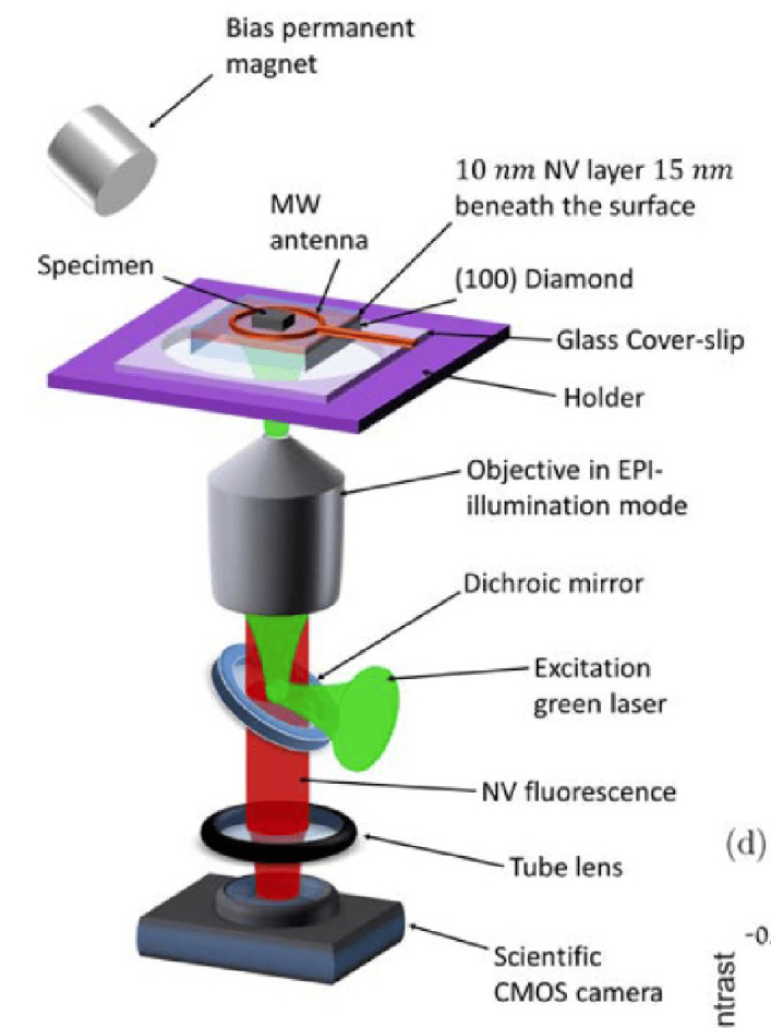
Figure 1: Custom-built Wide-field magnetic imaging microscope. The sample is placed on the surface of a diamond chip, which is implanted with a high-density thin layer of NV centres near the surface. The diamond is attached to the coverslip using immersion oil which is glued to the holder. Optical pumping green laser is incident through the bottom-polished side of the diamond surface using the objective in EPI mode. Coherent MW-field manipulation, which is created by an MW antenna, is located near the diamond surface containing the NV layer. The NV fluorescence passes through the diamond, the coverslip and the dichroic mirror and is then imaged onto a camera using the objective and a tube lens.
Project goals
- Optimize the diamond substrate, nanofabrication and NV integration.
- Learn and implement the MRI sensing techniques relevant for cellular MRI.
- Construct the system and perform initial measurements.
Supervision team
Professor Steven Prawer - The University of Melbourne
Professor Nir Bar-Gill and Professor Alex Retzker - Hebrew University of Jerusalem
Who we are looking for
Skills and requirements:
- Demonstrated experience in the field of experimental physics and/or engineering.
- Demonstrated ability to work independently and as part of the team.
- Demonstrated time and project management skills.
- Demonstrated ability to write research reports or other publications to a publishable standard (even if not published to date).
Further details
The PhD candidate will benefit from the combined expertise of the project supervisors, and the embedding into two research environments.
Professor Steven Prawer at the University of Melbourne will contribute expertise in nanofabrication and integrated photonics. Professor Nir Bar-Gill at the Hebrew University of Jerusalem will contribute expertise in quantum measurement.
The candidate will be enrolled in the PhD program in the School of Physics at the University of Melbourne and in the PhD program in the department of Physics at the Hebrew University of Jerusalem
To apply for this joint PhD opportunity, and to view the entry requirements, visit How to apply.
First published on 22 June 2022.
Share this article
Related items
-
How to apply
Apply for a joint PhD with the Jerusalem - Melbourne Joint PhD program.
-
Current projects
Discover what researchers from the Jerusalem - Melbourne Joint PhD are working on right now.
-
Your study experience
Discover what it's like to be a graduate researcher. Find out about University life, support services, and opportunities for skills development.
-
Find a supervisor or research project
Discover how to find a supervisor and learn how they can support your graduate research, or search available research projects.